The Oxygen Effect and Tumour Hypoxia: Essential Radiobiology for FRCR Part 1
Understand the oxygen enhancement ratio (OER), mechanisms of hypoxia-induced radioresistance, and strategies to overcome hypoxia for FRCR Part 1 radiobiology success.
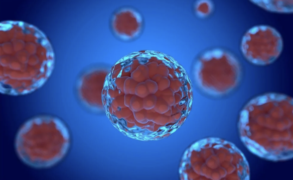
The oxygen effect is one of the most clinically important phenomena in radiobiology. Hypoxic tumour cells are significantly more resistant to radiation, impacting treatment outcomes. Understanding this effect is essential for FRCR Part 1 candidates.
Discovery and Significance
The oxygen effect was first observed in the 1920s and definitively demonstrated by Gray in 1953. He showed that well-oxygenated cells are approximately 2.5-3 times more sensitive to radiation than hypoxic cells. This has profound implications for radiotherapy, as solid tumours commonly contain hypoxic regions.
The Oxygen Enhancement Ratio (OER)
Definition: OER is the ratio of radiation doses required to produce the same biological effect under hypoxic versus aerobic conditions.
OER = Dose under hypoxia / Dose under normoxia (for same effect)
Typical OER Values:
- For low-LET radiation (X-rays, gamma rays): OER = 2.5-3.0 at high doses
- At clinical doses (~2 Gy per fraction): OER ≈ 2.0
- For high-LET radiation (alpha particles, heavy ions): OER approaches 1.0
This means hypoxic cells require 2-3 times the radiation dose to achieve the same cell kill as oxygenated cells.
Mechanism: The Oxygen Fixation Hypothesis
The most widely accepted explanation is the oxygen fixation hypothesis:
- Radiation creates free radicals (predominantly from water radiolysis)
- These radicals damage DNA, creating potentially lethal lesions
- In the presence of oxygen: O₂ "fixes" (makes permanent) the DNA damage by forming stable peroxy radicals
- In the absence of oxygen: Reducing agents (e.g., thiols like glutathione) can chemically restore the damaged DNA before the damage becomes permanent
Critically, oxygen must be present during irradiation or within milliseconds for the effect to occur - later oxygenation does not sensitise.
The Oxygen Effect Curve
Radiosensitivity increases rapidly as oxygen tension rises from 0, then plateaus:
- K-value (half-effect): ~3 mmHg (0.5% O₂) - the pO₂ at which radiosensitivity is midway between anoxic and fully aerobic
- Sensitivity is 90% of maximum at ~20 mmHg
- Further increases in pO₂ above ~20-30 mmHg provide minimal additional sensitisation
This is clinically relevant because:
- Normal tissue pO₂: 20-80 mmHg (fully radiosensitive)
- Tumour hypoxic regions: <10 mmHg (significantly radioresistant)
- Chronic hypoxia: <1-2 mmHg (maximally radioresistant)
Types of Tumour Hypoxia
1. Chronic (Diffusion-Limited) Hypoxia:
- Results from limited oxygen diffusion distance (~70-150 μm from vessels)
- Cells beyond this distance become chronically hypoxic
- Predictable, stable pattern
2. Acute (Perfusion-Limited/Cycling) Hypoxia:
- Results from temporary vessel closure or flow fluctuations
- Transient and unpredictable
- Cells may be hypoxic during one fraction but oxygenated during another
- Contributes to reoxygenation between fractions
LET and the Oxygen Effect
The OER decreases with increasing Linear Energy Transfer (LET):
- Low LET (X-rays, electrons): OER = 2.5-3.0
- Intermediate LET: OER progressively decreases
- High LET (alpha particles, heavy ions): OER ≈ 1.0-1.5
This is because high-LET radiation causes more direct DNA damage (independent of oxygen-mediated free radical damage) and creates such dense ionisation tracks that local oxygen is depleted.
This provides rationale for using high-LET radiation (carbon ions, protons at Bragg peak) for hypoxic tumours.
Strategies to Overcome Hypoxia
1. Fractionation and Reoxygenation:
- Reoxygenation is one of the 5 Rs of radiobiology
- Between fractions, previously hypoxic cells may become oxygenated as tumour shrinks and surviving hypoxic cells move closer to blood vessels
2. Hyperbaric Oxygen:
- Breathing oxygen at high pressure increases tissue oxygenation
- Shown to improve local control in head and neck cancers
- Logistically challenging
3. Hypoxic Cell Sensitisers:
- Nitroimidazoles (misonidazole, nimorazole) mimic oxygen's sensitising effect
- Nimorazole improved outcomes in head and neck cancer (DAHANCA trials)
4. Hypoxia-Activated Prodrugs:
- Drugs activated specifically in hypoxic environments
- Example: Tirapazamine
5. High-LET Radiation:
- Carbon ion therapy has reduced OER
- May be advantageous for radioresistant, hypoxic tumours
6. Modifying Oxygen Consumption:
- Metformin reduces mitochondrial oxygen consumption
- Increases tumour oxygenation in preclinical models
Clinical Implications
- Tumour hypoxia predicts poor prognosis across multiple cancer types
- Hypoxia imaging (FMISO-PET, pimonidazole) can identify hypoxic tumours
- Dose painting to hypoxic subvolumes is under investigation
- Anaemia should be corrected to optimise oxygen delivery
Key Exam Points
- OER = dose in hypoxia / dose in normoxia for same effect
- OER for low-LET radiation: 2.5-3.0 (at high doses); ~2.0 at 2 Gy
- OER decreases with increasing LET (approaches 1.0 for high LET)
- Oxygen fixation hypothesis: O₂ "fixes" free radical damage to DNA
- K-value (~3 mmHg): pO₂ at half-maximum sensitisation
- Chronic hypoxia: diffusion-limited; Acute hypoxia: perfusion-limited
- Reoxygenation between fractions partially overcomes hypoxia
- Nitroimidazoles are hypoxic cell sensitisers
Enjoyed this article?
Share it with a colleague preparing for their FRCR.